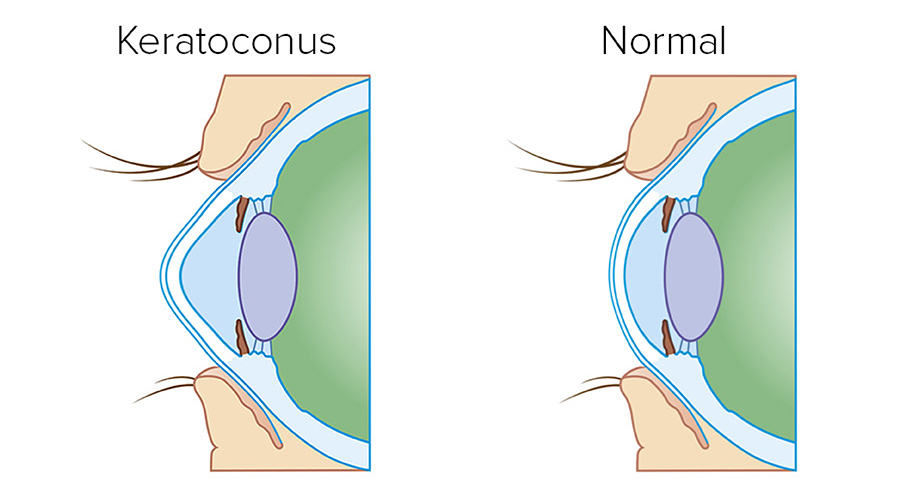
Keratoconus is a degenerative, hereditary corneal disease that usually occurs on both eyes. It is characterized by the cone-like bulge and central cornel thinning. Keratoconus usually occurs during puberty and about 20 percent of patients have the disease so advanced that surgical treatment (corneal transplantation) is necessary. Simultaneously with the corneal thinning and deformation, the diopter increases and the visual acuity decreases.

By applying semi-rigid contact lenses, an attempt to stabilize the deformed cornea, improve visual acuity and slow down the development of the disease is made. If the disease progresses, further thinning and deformity occur, and the scar-like changes in the central cornea begin to appear.
Cross linking is a procedure to strengthen the cornea and prevent further progression of keratoconus. Holding collagen fibers together is made possible using riboflavin solution and UVA radiation. Thus, satisfactory corneal mechanical strength is formed which helps to prevent further deformation and halt the progression of keratoconus.
Candidates for treating keratoconus with the cross-linking method are patients with keratoconus who are not older than 40 years of age and have no corneal scars. Prosječna debljina ljudske rožnice iznosi oko 550 mikrometara. Kod oboljelih od keratokonusa debljina je često ispod 500 mikrometara, a katkad i ispod 400 mikrometara. Dokle god se rožnica nije stanjila ispod 400 mikrometara, treba pokušati zaustaviti bolest primjenom Cross linking metode. To je nova metoda kojom se potiče umrežavanje kolagenih niti rožnice.

In order to use cross linking, no special preoperative preparation is needed, but a thorough ophthalmic examination and a cornea screening will be required which will show the actual state of deformation, i.e. keratoconus. Keratometric values (corneal curvature) above 50 microns are susceptible to keratoconus. A patient who is diagnosed with keratoconus - and is also prescribed for treatment - must undergo a complete ophthalmologic examination including visual acuity check-up, eye pressure measurement, biomicroscope scan, cornea topography and detailed posterior segment of the eye examination.
 Cross linking is a photooxidative method. Before the UVA radiation, anesthetic drops are applied and then the cornea epithelium is partially removed to enhance the riboflavin diffusion into the corneal tissue.
Thereafter, a riboflavin solution is applied every 2 minutes during 20 minutes. Riboflavin absorbs UV radiation and serves as a photosensitizer to create reactive oxygen. Ninety percent of UV light is absorbed in the cornea so there is no risk of lens or retina damage.
The length of radiation is between 10 and 30 minutes depending on which protocol is used during the procedure. If the patient is a younger person with keratoconus progressing rapidly, the so-called Dresden protocol is applied and the drip and UV radiation are carried out for 30 minutes. For the elderly and those with more stable keratoconus, shorter radiation is applied. The procedure is completely painless.
After the procedure a soft contact lens is placed on the cornea as a protection and left during complete regeneration of the epithelial cornea layer, usually for a few days. Antibiotic eye drops are applied for two to three weeks, as well as painkillers, as needed.
During the first days after surgery, a milder foreign body sensation may appear. The feeling of discomfort can be alleviated by applying artificial tears. After the procedure, the necessary follow-up examinations are required. As a rule, two to three check-ups are required in the first month after the operation. It is necessary to wear sunglasses as a protection when you go out. All daily activities are allowed, but during the first few weeks, dust, wind and smoke-filled rooms should be avoided. Visual acuity gradually recovers. One month after the operation it is possible to continue wearing semi-rigid contact lenses. It is important to emphasize that this method will not improve your vision, but will stabilize it to so that the cornea can become thinner even further at least in the first phase. The most important thing is to stop the disease in its development, and the effect of the operation lasts up to 10 years. During follow-up examinations the visual acuity and condition of the cornea are measured by corneal topography 2x every year for the next few years.
Serious complications after keratoconus surgery using cross linking method have not been known so far. As transient side effects during the first weeks after surgery, redness of the eye, itching, choking and blurred vision may occur.
Become a part of the Polyclinic Bilić Vision family of satisfied patients and call today to be informed of keratoconus treatment using cross linking method. You can make an appointment over the phone: + 385 1/4678-444 or by email at info@bilicvision.hr.
Need more information? Can't find what you are looking for or want to make a doctor's appointment? Leave your information and we will contact you as soon as possible:
Vaš upit je zaprimljen! Hvala Vam na povjerenju!